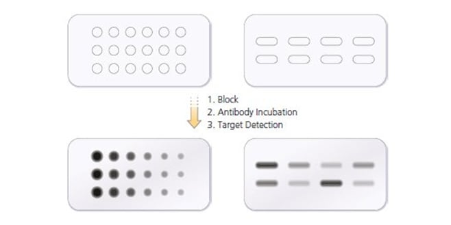

Representative images of OB* incubated for 24 h were also shown as positive control. b Representative confocal microscope images showing SF (at 0.3 muM) incubated in CM without cells in wells containing a glass coverslip for 0-24 h at 37 degC. a Dot-blot analysis of alphaS species probed with conformational specific antibody A11 (AHB0052, Thermo Fisher Scientific), OC (AB2286, Sigma-Aldrich), conformation-insensitive polyclonal anti-alphaS antibody (ab52168, Abcam) and conformation-insensitive monoclonal 211 antibody specific for human alphaS (sc12767, Santa Cruz Biotechnology). 4 alphaS fibrils gradually release oligomers that are ultimately responsible for their toxicity. k Abeta plaques in the cerebral cortex of anįig.

j Representative example of a retinal wholemount on which 6E10-positive Abeta plaques are depicted with dots to illustrate plaque distribution in the retina. f Example of a reconstruction of the z-plane of the image in panel e, with 82E1 and DAPI nuclear counterstaining, showing that Abeta plaques mainly localize to the inner plexiform layer. d - i Abeta immunostainings with antibodies 82E1 ( d - f ) and 6E10 ( g - i ) on retinal wholemounts of 18-month-old WT ( d, g ) and App NL - G - F mice ( e, f, h, i ) reveal the presence of Abeta plaques in App NL - G - F mice only. Of note, human Abeta levels are undetectable in the retinas of WT mice and therefore these controls are not shown. c Eighteen-month-old APP/PS1 mice have comparable soluble Abeta 42 levels yet lower levels of insoluble Abeta 42 and higher soluble Abeta 40 than App NL - G - F mice of the same age. One-way ANOVA with Dunnett's multiple comparisons test (F 5,28 = 6.686, p = 0.0006 for soluble Abeta 42 F 5,27 = 20.27, p < 0.0001 for insoluble Abeta 42 ) n = 5-6. Accumulation of soluble, but not insoluble, Abeta 40 is seen at all ages yet does not progress with age. Levels of insoluble Abeta 42 only start to rise at 12 months. a, b Amyloid ELISA on retinal lysates of App NL - G - F mice shows accumulation of soluble Abeta 42 from 3 months of age, which rises up till 24 months. 1 Amyloid burden increases with age in the retina of App NL - G - F mice.


 0 kommentar(er)
